- Micro-computed tomography (Micro-CT) service
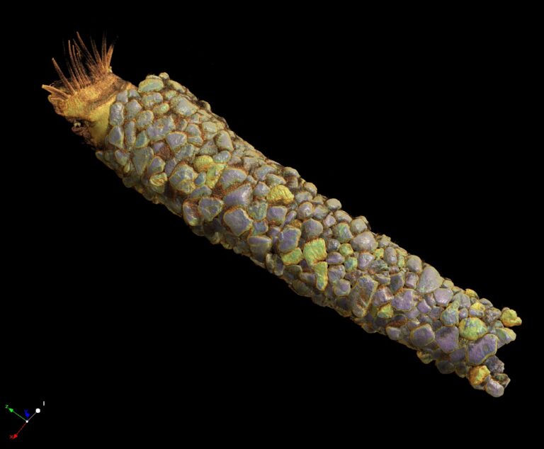
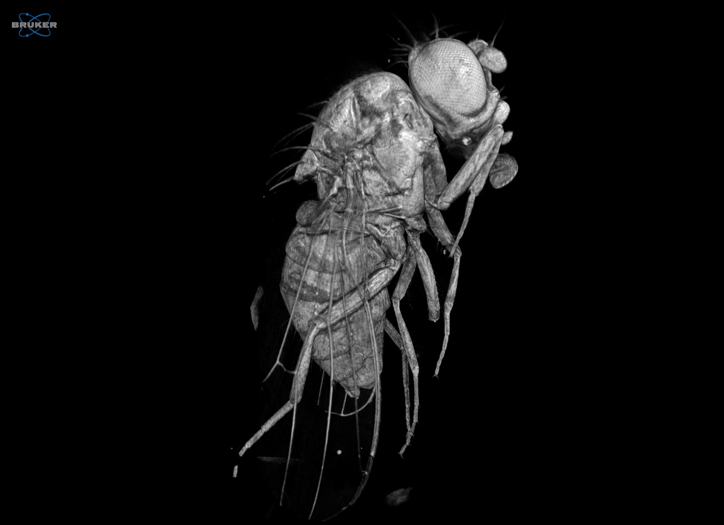
Micro-CT imaging services are offered using a Skyscan 1172 (Bruker, Belgium) microtomograph, which is a non-destructive imaging technique, based on X-Rays.
This service allows the creation of high-resolution three-dimensional data and includes specimen preparation, scanning, reconstruction, 3D volume rendering and 3D analysis.
The scanner uses a tungsten X-ray source with an anode voltage ranging from 20 to 100 kV, 11 MP CCD camera (4000 × 2672 pixel) and a maximal resolution of 0.8 μm/pixel. The maximum object size that can be scanned is 50mm in diameter. The scanning duration depends on the sample size and the selected scanning parameters (resolution, averaging frames, etc).
- Micro-CT virtual laboratory (Micro-CTvlab )
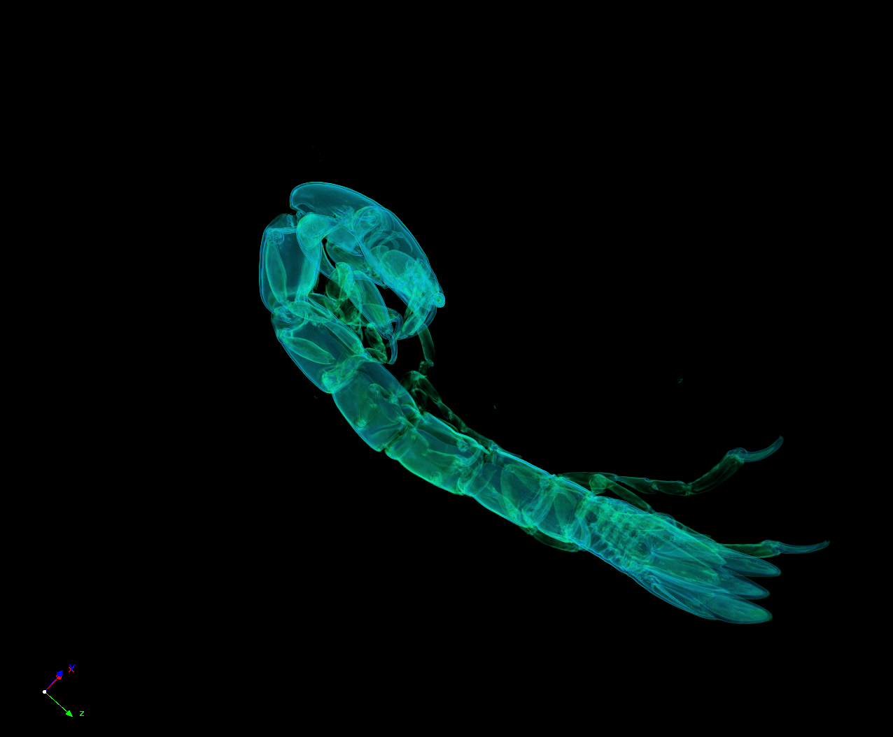
The virtual micro-CT laboratory (Micro-CTvlab), supported by the ESFRI LifeWatchGreece infrastructure, offers a collection of virtual image galleries of various biological materials which can be displayed and disseminated through a web-based framework. This virtual lab allows the user to manipulate the 3D models through a series of online tools or to download the datasets for local manipulations. The data and functions of the Micro-CTvlab can be accessed either on a normal computer or through a dedicated version for mobile devices.
- BIOIMAGING virtual laboratory
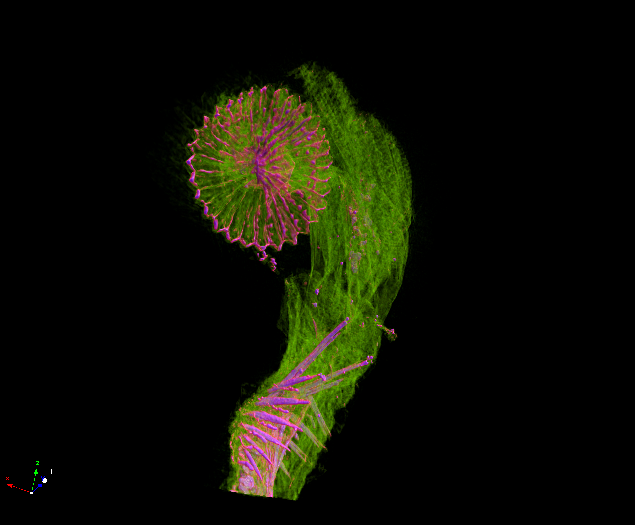
The BIOIMAGING virtual lab (under development) was created during BioImaging-GR in order to display and disseminate virtual galleries of various medical and biological materials. This virtual lab offers a collection of images generated by several imaging technologies (e.g. electron microscopy, fluorescence microscopy etc.). Furthermore, the BIOIMAGING virtual lab is linked with the Micro-CTvlab in which 3D virtual samples are presented and can be interactively displayed and retrieved through a web-based application. BIOIMAGING virtual lab is also linked with the AQUAPATHvlab which is a virtual fish pathology laboratory supported by the MOUNT (MOdern UNifying Trends in marine biology) project, and it offers a collection of several imaging techniques, including microtomography, histology, SEM, TEM that can be used for disease diagnostics.